Science-- there's something for everyone

Tuesday, December 11, 2012

You’re always on Mom’s mind


Researchers, led by William Chan, currently at the University of Alberta, have found fetal cells in the brains of mothers. Yes, kids, you really are always on our minds.

I’ve written before about a phenomenon called ‘fetal microchimerism’. The tissues of any woman who has ever been pregnant, regardless of whether that pregnancy resulted in a live birth, contain a few cells from the fetus or fetuses she was carrying. For better or worse, these fetal cells remain in the mother for the rest of her life. They’ve been associated both with tissue repair and protection from cancer and with the initiation of autoimmune diseases. It hasn’t been clear, until now, whether the fetal cells can also make their way into women’s brains.

The easiest way to distinguish maternal from fetal cells is to screen for cells that have a Y chromosome, which will only be present in the cells of any male fetus they carry. This is exactly what the researchers did. They performed brain autopsies on 59 women (age 32 to 101 at time of death) and tested up to twelve regions within each brain for the presence of Y chromosomes. 

Around 60% of the women had Y carrying fetal cells in their brains, and nearly all brain regions were affected. Clearly, fetal cells are crossing the blood-brain barrier and making homes for themselves in their mother’s brains. Remember, these experiments were conducted many decades after the pregnancies that gave rise to these cells. I’d also like to point out that the numbers of fetal cells would naturally have been much higher if the researchers could have screened for female fetal cells as well as male ones. It could be that the entire brain of any woman who has ever been pregnant is permeated with cells from all those fetuses. Even more interesting, since maternal cells pass into the infant during breastfeeding, it could be that a few of those cells are from prior siblings. We could all be multiple microchimeras.

The authors also investigated whether having this fetal microchimerism could affect whether women went on to suffer from Alzheimer’s disease (AD). Although they found that women who had suffered from AD were less likely to test positive for male fetal cells, this result was not significant. A far larger study will be needed before any general conclusions can be drawn about this.

 Story submitted by Cathy Earle.

Chan WF, Gurnot C, Montine TJ, Sonnen JA, Guthrie KA, & Nelson JL (2012). Male microchimerism in the human female brain. PloS one, 7 (9) PMID: 23049819.


Monday, December 10, 2012

Bad news for coastal cities


An international team of scientists, led by Andrew Shepherd of the University of Leeds, has put together the most comprehensive study of Arctic and Antarctic ice sheet loss to date. They also determined how much that loss of ice is affecting global sea levels. The news is not good.

The 47 authors used nineteen years of satellite data to come to their conclusions. They found that the loss of the Antarctic and Greenland ice sheets have contributed to a rise in sea level of eleven millimeters over the past two decades. While this may not sound like a lot, remember, that rise is continuing and increasing. Both poles are losing ice at a faster pace. As Erik Ivins from NASA’s Jet Propulsion Laboratory points out:
The rate of ice loss from Greenland has increased almost five-fold since the mid-1990s.
This means that the rise of the oceans due solely to polar ice sheet loss has increased to nearly a full millimeter per year. However, the bulk of the sea level rise is caused not by the added water from the melting ice but by the expanding of the ever warmer water. In other words, global warming will continue to be a problem even after the polar ice caps have already disappeared.

You can see an interview with Andrew Shepherd and Erik Ivins below. To watch an animation of the researchers' results, click here.


Shepherd, A., Ivins, E., A, G., Barletta, V., Bentley, M., Bettadpur, S., Briggs, K., Bromwich, D., Forsberg, R., Galin, N., Horwath, M., Jacobs, S., Joughin, I., King, M., Lenaerts, J., Li, J., Ligtenberg, S., Luckman, A., Luthcke, S., McMillan, M., Meister, R., Milne, G., Mouginot, J., Muir, A., Nicolas, J., Paden, J., Payne, A., Pritchard, H., Rignot, E., Rott, H., Sorensen, L., Scambos, T., Scheuchl, B., Schrama, E., Smith, B., Sundal, A., van Angelen, J., van de Berg, W., van den Broeke, M., Vaughan, D., Velicogna, I., Wahr, J., Whitehouse, P., Wingham, D., Yi, D., Young, D., & Zwally, H. (2012). A Reconciled Estimate of Ice-Sheet Mass Balance Science, 338 (6111), 1183-1189 DOI: 10.1126/science.1228102


Friday, December 7, 2012

A new contraceptive


University of Washington researchers, led by Cameron Ball, have invented a new method of contraception. Best of all, their device, made of drug-eluting nanofibers, blocks not only sperm but also sexually transmitted diseases (STDs) like HIV.

Contraceptives and STD preventatives can be classed into two broad categories depending on their mode of action. There are chemical barriers, which can contain hormones, spermicides and/or microbicides. These compounds can be administered as pills, gels, intravaginal rings or vaginal films. There are also physical barriers, the best known of which is the condom. Ideally, a contraceptive would employ both chemical and physical barriers to block sperm and infectious agents. That’s exactly what these new nanofibers do.

The researchers used electrospinning to create their fiber meshes out of materials that are already approved for use in human implants. They used a tampon applicator as a template to ensure easy installation, and incorporated a variety of anti-HIV agents into the finished product. The end result is a soft, flexible object that releases one or more antiviral drugs.

In one of their tests, the scientists incorporated a dye into the fibers and inserted the mesh devices into female mice. Within 30 minutes, the mice’s vaginal tracts were completely coated with dye, indicating that the fibers had indeed released their drug load. When tested in tissue culture experiments, the mesh devices could inhibit both sperm motility and HIV transmission, depending on which drugs they contained. In addition, the fibers present an impenetrable barrier to sperm.


Caption: The electrospun fibers can release chemicals or, as shown here, they can physically block sperm.
Credit: Kim Woodrow, Univ. of Washington.

The invention fulfills the five criteria that Ball and his colleagues set for themselves. The fibers are inexpensive, can deliver multiple drugs, are fully reversible, provide extensive coverage of mucosal tissue and can be managed solely and discreetly by women. This last is important because women can’t always control what their partners are willing to use. 

The next step is to fine-tune the drug delivery system. Different raw materials and thicknesses can determine how quickly the drugs are dissipated throughout the vagina. Thus, it may be possible to create devices for both long term (sustained release) and immediate (quick dispersal) usage, depending on the woman’s needs. And after all, the more choices women have for protecting their health and controlling their fertility, the better.


Cameron Ball, Emily Krogstad, Thanyanan Chaowanachan, & Kim A. Woodrow (2012). Drug-Eluting Fibers for HIV-1 Inhibition and Contraception PloS ONE : doi:10.1371/journal.pone.0049792.


Thursday, December 6, 2012

Detecting breast cancer via methylation levels


Epigenetics, and specifically DNA methylation, is in the news quite a lot. I’ve explained DNA methylation before (here and here). Suffice it to say that this ‘after-factory’ alteration does not affect the sequence of DNA but does affect the expression of that DNA, often by inactivating the genes involved. However, you may be asking, ‘when is this new field of study going to yield anything useful?’ If you or someone you know is at risk for developing breast cancer, that day may be sooner than you think.

Researchers from various German universities and research hospitals compared methylation levels in ten breast cancer tumors (two from pre-invasive tumors and eight from invasive cancers) and ten normal controls. Overall, they found over 200 sites that were significantly hypermethylated in the tumor samples. In particular, they found nine genes that were already significantly hypermethylated in the pre-invasive tumors. In fact, methylation levels did not differ much between pre-invasive and invasive tumors or between different tumor subtypes. This finding suggests that methylation can be used as a tool for the early detection of breast cancer tumors.

If you consider that the five-year survival rate for breast cancer can increase to nearly 100% (depending on the type of cancer) if the disease is detected early enough, the importance of this finding cannot be overstated. The next step is to figure out whether the extreme methylation is a cause or an effect of the tumorigenesis. Interestingly, many of the genes that are hypermethylated, and thus inactivated, in breast cancer samples are homeobox genes (genes that are involved in embryonic development). The authors speculate that this may indicate that the breast cancer tumor cells originated as stem cells.


Faryna, M., Konermann, C., Aulmann, S., Bermejo, J., Brugger, M., Diederichs, S., Rom, J., Weichenhan, D., Claus, R., Rehli, M., Schirmacher, P., Sinn, H., Plass, C., & Gerhauser, C. (2012). Genome-wide methylation screen in low-grade breast cancer identifies novel epigenetically altered genes as potential biomarkers for tumor diagnosis The FASEB Journal DOI: 10.1096/fj.12-209502


Wednesday, December 5, 2012

Just for fun: Dew-covered insects


David Chambon had captured some amazing photographs of insects bejeweled with dew. Here's one example, but definitely go to his website to see the rest.

Macro Photographs of Dew Covered Dragonflies and Other Insects by David Chambon macro insects


Tuesday, December 4, 2012

Bad news for cleaning up oil spills


Just when you thought the 2010 oil spill in the Gulf of Mexico couldn’t have been worse. Roberto Rico-MartĆ­nez from Universidad Autónoma de Aguascalientes and Terry Snell and Tonya Shearer from the Georgia Institute of Technology have found that the very oil dispersants used to ameliorate the problem could have made it fifty times worse.

As test subjects, the scientists chose five strains of Brachionus plicatilis. Not only are these rotifers (tiny marine organisms found in zooplankton) a critical part of the marine food chain, but they are also notoriously sensitive to environmental conditions.

The researchers exposed the rotifers to varying amounts of crude oil and/or Corexit 9500A oil dispersant (a product used in the Gulf). The LC50 (lethal concentration 50, or amount of product that will kill 50% of the organisms) for each combination was determined. Depending on the strain of B. plicatilis tested, the LC50 with crude oil alone was between 2 and 20 mg oil/liter water. Oil dispersant alone yielded LC50s ranging from 0.5 to 14 mg/L. When the two components were mixed in ratios of 1:10 or 1:50 dispersant to oil (the recommended dosages), the LC50 decreased to 0.2 to 0.3 mg/L. In other words, the combination of crude oil plus oil dispersant is far more toxic to rotifers than either chemical alone.

There was one glimmer of good news. When the researchers tested a ratio of 1:130 dispersant to oil (the conditions actually used at the Deep Water Horizon disaster), toxicity did not increase compared to using either component alone. This makes sense since the 1:50 ratio was slightly less toxic than the 1:10 ratio. Of course, you’re never going to find a ratio that’s less toxic than the dispersant alone, which is pretty darned lethal all by itself. Thus, the best you’re going to do is to not make things worse. Not exactly a glowing endorsement for a rescue technique.

Unfortunately, I think this means it’s back to the drawing board for cleaning up oil spills.

Rico-MartĆ­nez R, Snell TW, & Shearer TL (2012). Synergistic toxicity of Macondo crude oil and dispersant Corexit 9500A(®) to the Brachionus plicatilis species complex (Rotifera). Environmental pollution (Barking, Essex : 1987), 173C, 5-10 PMID: 23195520


Monday, December 3, 2012

Correcting trisomy in cells


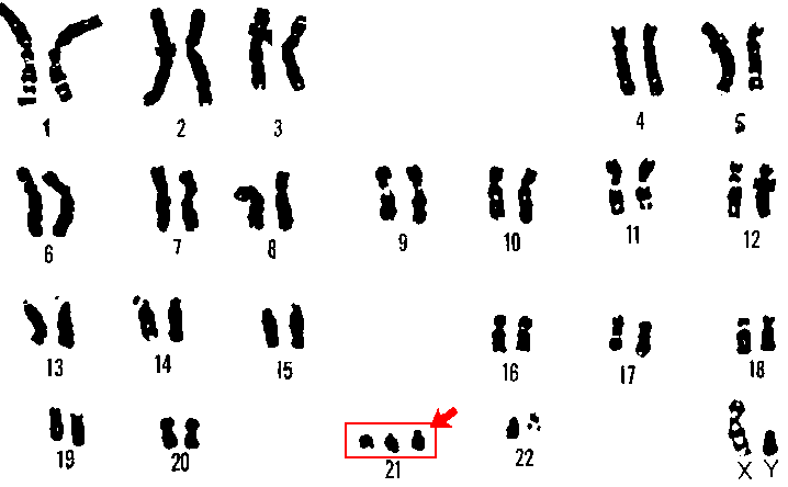
We’re all supposed to have two of each chromosome (unless you’re a boy, in which case you have one mismatched pair of sex chromosomes). Having three of one chromosome (trisomy) is usually lethal in uteru, but can sometimes only cause the kind of severe health and cognitive problems seen in people with Down syndrome (the result of having three chromosome 21s, shown below). What if we could pluck one of those extraneous chromosomes right out of a cell? Thanks to the work of researchers led by Li Li of the University of Washington, Seattle, we’re one step closer to that goal.


The researchers generated some induced pluripotent stem cells with trisomy 21. One of those three chromosome 21s was genetically engineered to include a gene that was detrimental to the cell under certain conditions. Thus, the scientists were able to put selective pressure on the cells to lose that extra copy of chromosome 21. This is precisely what happened. Three quarters of the surviving cells had shed the malignant chromosome.

Obviously, we’re still far from being able to remove every extra chromosome in a Down syndrome patient’s body. However, we may be able to modify this technique to remove chromosomes in a few critical cells and then reintroduce them back into the person’s body. In addition, we may be able to rid stem cell cultures of the trisomies that frequently plague them. Either way, this method will probably continue to be used by molecular biologists.


Li, L., Chang, K., Wang, P., Hirata, R., Papayannopoulou, T., & Russell, D. (2012). Trisomy Correction in Down Syndrome Induced Pluripotent Stem Cells Cell Stem Cell, 11 (5), 615-619 DOI: 10.1016/j.stem.2012.08.004