DNA is subject to damage from UV light, toxins, and misreading during replication. These errors are repaired by a variety of specialized proteins that check and fix the DNA strands. Researchers led by Bennett Van Houten of the University of Pittsburgh have been able to directly observe this process in E. coli.
They labeled two exinuclease repair proteins, UvrA and UvrB with different colored quantum dots (semi-conductor nanocrystals). They combined the tagged proteins with untangled stretches of DNA they called ‘tightropes’ and watched what happened.

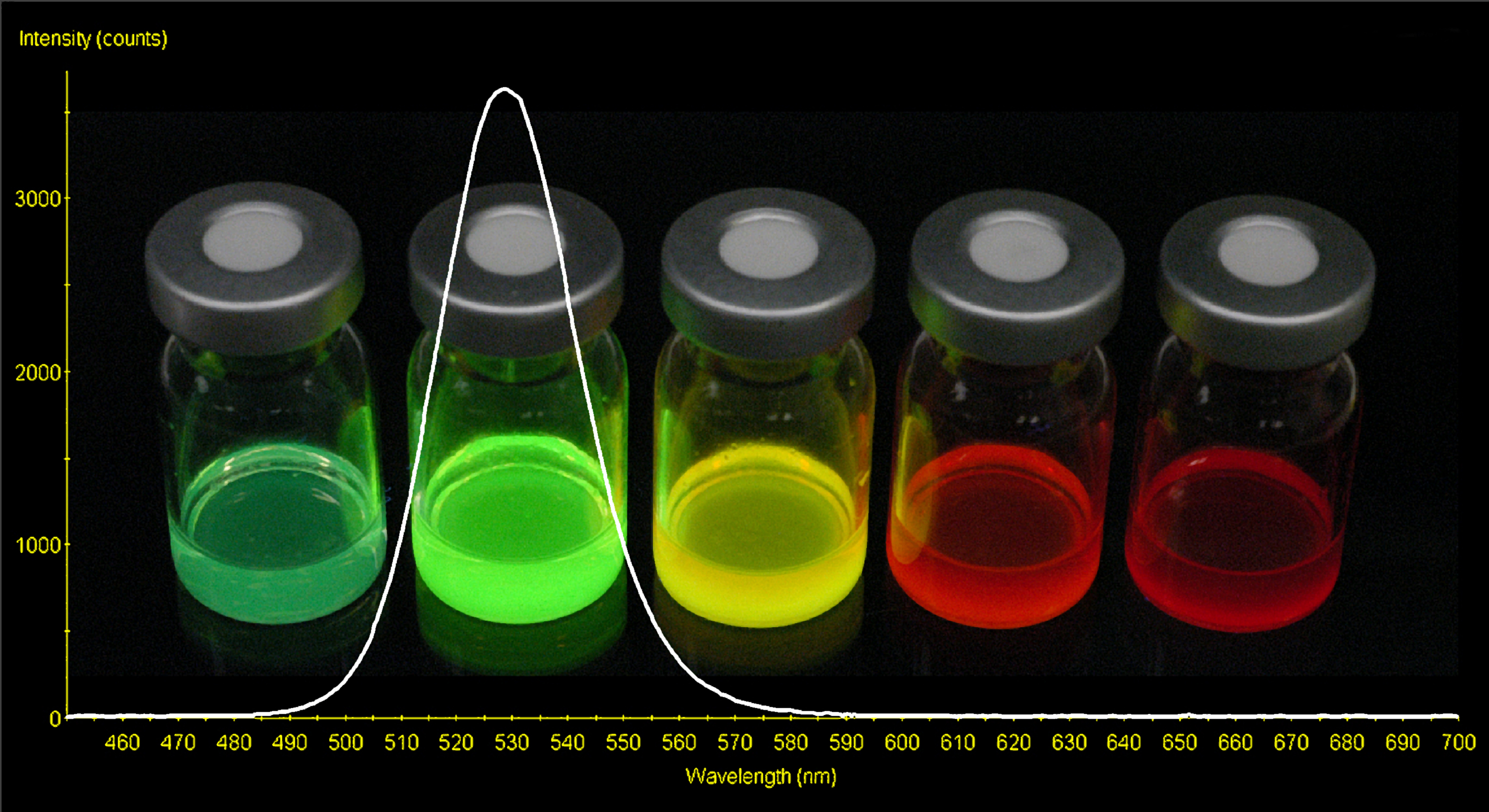
Cadmium selenium Quantum Dots (metal nanoparticles that fluoresce in a variety of colors determined by their size)
UvrA alone would bind to a spot on a DNA molecule, remain in place for about seven seconds, then hop to a different DNA molecule. UvrB alone did not bind DNA at all. When UvrA was combined with UvrB, this new complex was capable of sliding along the DNA strand for up to forty seconds. About a third of the UvrAB complexes exhibited what the scientists called ‘paused motion’, in which the repair proteins seemed to hesitate at specific sites.
Co-author David Warshaw of the University of Vermont suggested:
Paused motion could represent UvrAB complexes checking for structural abnormalities associated with DNA damage.
I dealt with the issue of proteins moving along DNA strands in a previous post, though at that time, the interaction was not directly observed.
No comments:
Post a Comment