There are two ways in which cells die. The first is called apoptosis, or programmed cell death. In multicellular organisms, apoptosis kills isolated cells and is often a necessary part of development. For example, the webbing between our fingers is removed during embryogenesis by apoptosis. The other type of cell death is caused by necrosis. Unlike apoptosis, necrosis is not a normal feature of the cell life cycle. It results from injury (trauma, infection, toxins, etc) and can cause whole tissues or entire organisms to die. Necrotic cell death is characterized by calcium intake and other factors.

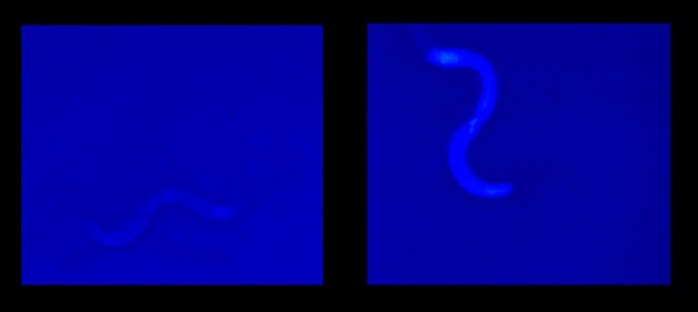
C. Elegans alive (left) and dead (right).
Where does C. elegans come in? While normally transparent, C. elegans cells are kind enough to fluoresce bright blue when they die. The authors call this ‘death fluorescence’. Older worms have more of these fluorescent particles than younger worms do. You can tell how close a worm is to the end of its natural life by measuring that fluorescence. However, regardless of age, each worm glows much more brightly at the time of death. Even more intriguing (or morbid, depending on your sensibilities), you can watch a blue wave of death propagate throughout the dying worm.
You can watch a worm turn bright blue and die below.
There is a bright side to all this, even for the worms. When scientists created conditions that would normally kill worms (such as exposing them to heat or cold), they were able to stop the death progression by blocking the necrotic pathway. This only worked in stress-related death though. Blocking necrosis did not help worms who were dying of old age.
Although they don’t turn blue as they die, mammalian cells have similar necrotic pathways. Thus, studying death fluorescence in nematode worms could lead to insights into how more complex creatures progress from life into death.
No comments:
Post a Comment