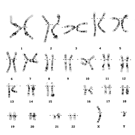
You’re probably used to thinking of chromosomes as looking like this:

 |
This is the chromosome structure from single-cell Hi-C.
Credit: Dr. Peret Fraser, Babraham Institute.
The researchers used a technique called ‘chromosome conformation capture’ (Hi-C) to combine data from thousands of molecular measurements within single cells. Not only could this method be used to visualize the DNA in non-dividing cells, but also to see how chromosomes interact with each other. In addition, it could help scientists ‘see’ which genes are active, based on their positioning.
Douglas Kell, Chief Executive of Biotechnology and Biological Sciences Research Council explains:
Until now, our understanding of chromosome structure has been limited to rather fuzzy pictures, alongside diagrams of the all too familiar X-shape seen before cell division. These truer pictures help us to understand more about what chromosomes look like in the majority of cells in our bodies. The intricate folds help to unravel how chromosomes interact and how genome functions are controlled.
No comments:
Post a Comment